






Hearing that a doctor wants to look into a probable malignancy or cancer might make a person and his/ her family worried. A blood test may be followed by a scan, a biopsy, and occasionally additional molecular or genetic testing. In this matter better guidance can be provided by an oncologist in Siligurior any place else.
Among different cancer diagnostic tests by doctors, some may seek abnormalities, others show where they are, some look at cells under a microscope, and others reveal biological or genetic traits that may affect treatment.
A single test may not detect everything about cancer. A biopsy is mostly required to verify the presence of malignancy. The 12 common tests that doctors may use to diagnose cancer are explained in this guide; have a read.
Why Do Doctors Order Cancer Diagnostic Tests?
An unexplained lump, a persistent cough, unexpected bleeding, unexplained weight loss, changes in bowel or bladder habits, or difficulty swallowing are some of the classic symptoms of cancer.
However, non-cancerous illnesses might also cause identical symptoms; therefore, a clinical diagnosis is vital. For this, medical professionals integrate data from a patient's medical history, physical examination, laboratory testing, imaging, pathology, and occasionally molecular testing.
A normal test result may not usually rule out cancer, nor does an abnormal test necessarily indicate one has cancer.
The goal is to answer several questions :
- Is there anything unusual that needs to be looked into?
- Is cancer genuinely present?
- What kind of cancer is it?
- Where did it start?
- Has it spread?
- What biological characteristics does it possess?
- Which therapies would be suitable?
With that framework in mind, here are 12 tests that may form part of a cancer diagnostic workup.
1. Complete Blood Count (CBC) and Other Blood Tests
Red blood cells, white blood cells, platelets, hemoglobin, and other blood aspects are measured during a complete blood count (CBC). Major anomalies may pinpoint anemia or other issues that need to be looked into, as well as giving hints about some blood malignancies, such as leukemia.
Tests for blood chemistry can evaluate proteins, electrolytes, kidney and liver function, and other elements. Doctors can use this data to assess general health and spot anomalies that could require more testing.
2. Urine Tests and Urine Cytology
When medical professionals suspect a kidney, bladder, or urinary tract issue, urine tests may be helpful. Blood, protein, or other anomalies may be seen in a standard urine test.
Urine cytology goes one step further by looking for abnormal cells that have been expelled from the urinary tract under a microscope. When looking at potential urinary cancers/ malignancies, doctors may combine urine tests with imaging or procedures like cystoscopy.
3. Mammography
Low-dose X-rays are used in mammograms to produce finely detailed images of breast tissue, and it can be used as part of screening.
Doctors may suggest more cancer diagnostic tests like imaging, such as imaging (MRI or ultrasound) and possibly a biopsy based on the results, besides a mammogram. This includes whether there are anomalies in the breast that need more examination, such as lumps, calcifications, or other changes.
4. Ultrasound
Ultrasound generates images of internal body structures using high-frequency sound waves, an ideal test for cancer diagnosis. Unlike X-rays and CT scans, normal ultrasonography does not use ionizing radiation; all are effective.
It is effective for checking the breast, thyroid, liver, kidneys, pelvic organs, testes, and lymph nodes; ultrasound is also used to guide a needle during some biopsies.
Because ultrasound can identify fluid-filled structures from many solid abnormalities, it can assist clinicians in determining what type of study to conduct next.
5. CT Scan
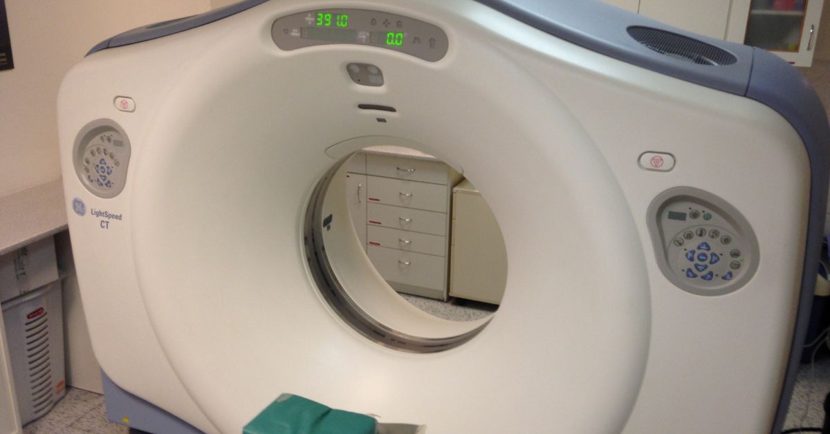
A computed tomography (CT) scan creates detailed cross-sectional images of the body by combining multiple X-ray images with computer processing. When checking the chest, abdomen, and pelvis, CT is generally very helpful.
It can be used by medical professionals to detect a suspicious lump, evaluate its location and size, inspect surrounding lymph nodes, or check for potential dissemination to other body areas. Sometimes specific structures can be easier to perceive by using contrast material.
6. MRI Scan
Magnetic resonance imaging (MRI) creates incredibly detailed images of soft tissues, and this is done by using radio waves and magnetic fields.
When clinical experts need a deeper look at regions including the brain, spinal cord, prostate, liver, pelvic, and other soft tissues, they recommend MRI.
To enhance visualization, contrast may occasionally be used. Like other imaging procedures, magnetic resonance imaging (MRI) is strong, but it typically detects anomalies rather than conclusively showing that an abnormality is cancer.
7. PET Scan or PET-CT
Insights into biological activity within the body can be obtained by a positron emission tomography (PET) scan. There are more radioactive glucose-like tracers.
Often, PET is used with CT to create PET-CT, which enables medical professionals to view both anatomical features and metabolic activity. In certain tumors, PET helps determine the extent of the disease, find potential metastases, measure the effectiveness of treatment, or look into a probable recurrence.
8. Endoscopy
Using a small, flexible device with a camera, endoscopy enables medical experts to examine directly within some body parts.
Examples include:
- Examining the colon and rectum is known as a colonoscopy
- The esophagus and stomach are examined during an upper endoscopy or gastroscopy
- A bronchoscopy looks at the airways
- The bladder is examined during a cystoscopy
One major benefit is that if a worrisome area is noticed, surgeons can collect a tissue sample during the same treatment.
9. Biopsy
A biopsy involves a surgeon removing tissue or cells from a location that appears suspicious. After that, a pathologist looks at the material under a microscope and might do more tests in the lab.
Based on the site, a biopsy could include:
- Fine-needle aspiration
- Incisional biopsy
- Excisional or surgical biopsy
- Core needle biopsy
- Endoscopic biopsy
- Bone marrow biopsy in some cases
Tissue examination is the gold standard for diagnosing many solid tumors. The kind, grade, and other treatment-relevant features of the malignancy may also be identified in the pathology report.
10. Tumor Marker Tests
Chemicals present in blood, urine, tissue, or other bodily fluids that may reveal details about certain tumors are known as tumor markers. PSA, CA-125, CEA, AFP, and CA 19-9 are a few examples.
The fact that tumor markers are mostly insufficiently accurate to identify cancer on their own is crucial. While some cancer patients may have normal marker levels, benign illnesses might occasionally boost them.
11. Genetic and Molecular Testing
Cancer is fundamentally a disease involving changes in cells, and some of those changes can be recognized through molecular testing. Medical experts may examine tumor tissue for specific genetic changes, proteins, hormone receptors, or other biomarkers.
Testing may search for changes related to BRCA, EGFR, KRAS, HER2, mismatch-repair deficiency, microsatellite instability, or other clinically significant biomarkers, based on the type of cancer. Differentiating between hereditary genetic testing and malignant genetic testing is also of great importance.
12. Cell-Based Tests and Specialized Pathology
Following the collection of tissue or blood, labs may carry out tests beyond standard microscopic inspection. These can involve immunohistochemistry, immunophenotyping, cytogenetic testing, and other specialist pathology procedures.
They are especially vital for diagnosing specific blood malignancies and differentiating cancer subtypes. For instance, cytogenetic analysis can uncover some chromosomal changes linked to specific diseases, whereas immunophenotyping can assist in identifying aberrant cell types in leukemia and lymphoma.
How Doctors Put Cancer Test Results Together
If a suspected abnormality is discovered, a doctor may suggest a biopsy. This is after starting with the symptoms and examination results and moving on to blood testing or imaging. Further tests may rule out the type, grade, biomarkers, and stage of cancer once it has been proven.
Staging poses a separate inquiry from diagnosis, such as how far the cancer has spread. The effective TNM system, the main tumor, adjacent lymph nodes, and distant metastases, is considered when staging various cancers.
To determine the severity of cancer, further imaging, laboratory testing, and treatments will be of great help.
How Should the Results of Your Cancer Test Be Interpreted?
It can be easy to believe that a suspicious scan indicates cancer or to look up the meaning of every highlighted figure on a lab report. Neither method is trustworthy.
Now, it is necessary to evaluate test results contextually. While a normal test might not entirely rule out cancer, a result outside of the reference range might have a non-cancerous reason. Along with symptoms, examination results, pathology, and other studies, imaging results must be considered.
You might receive expert guidance from Dr. Manish Goswami, offering fully patient-centric and comprehensive head & neck cancer care.
Final Words
The present approach to cancer diagnosis is not exclusive to a single test. Symptoms, physical examination, laboratory testing, imaging, endoscopy, biopsy, pathology, tumour markers, and molecular information are factors that determine the particular clinical situation.
A scan may reveal a mass, a blood test may cause alarm, and a tumour marker may be elevated, but the right mix of tests is what helps doctors achieve diagnostic accuracy.
FAQs
1. Which test is the most reliable for identifying cancer?
For every malignancy, there isn't a single test that is the most accurate, but to confirm the diagnosis of many solid tumours, a biopsy and pathological investigation are required.
2. Can cancer be detected by a blood test?
Some blood-based diagnostics are helpful for specific cancers, and some blood tests can offer hints. Plus, specific scenarios can also provide information markers. Nevertheless, most blood tests are unable to independently confirm or rule out malignancy/ cancer.
3. Can a CT scan or an MRI confirm cancer?
CT and MRI scans can detect abnormal masses and provide detailed insights into their size, location, and extent. However, an imaging abnormality may not necessarily indicate cancer, and a biopsy may be required for a conclusive diagnosis.
4. Are tumour markers sufficient to diagnose cancer?
Tumour markers can be raised due to cancer or some non-cancerous illnesses, but some cancer patients have normal marker levels. Cancer doctors evaluate tumour markers alongside other clinical and diagnostic findings.
5. Why is genetic testing carried out following a diagnosis of cancer?
Tumour features that could help categorize the cancer or direct treatment can be found by genetic or molecular testing. A person may be able to determine whether they have genetic changes associated with an increased likelihood of cancer through separate inherited genetic testing.
6. Does an abnormal cancer test result always mean I have cancer?
There may be non-cancerous reasons for abnormal blood tests, scans, tumour markers, or other results. In other instances, cancer is not always ruled out by a normal result. To make a diagnosis, cancer doctors may require multiple pieces of evidence and perhaps a biopsy.